
The x-ray machine will be placed over the lower part of your spine. A slipped disc can produce varying degrees of pain in the back or neck along with numbness or weakness in the corresponding organs arms or legs as follows.

Pin On Yoga
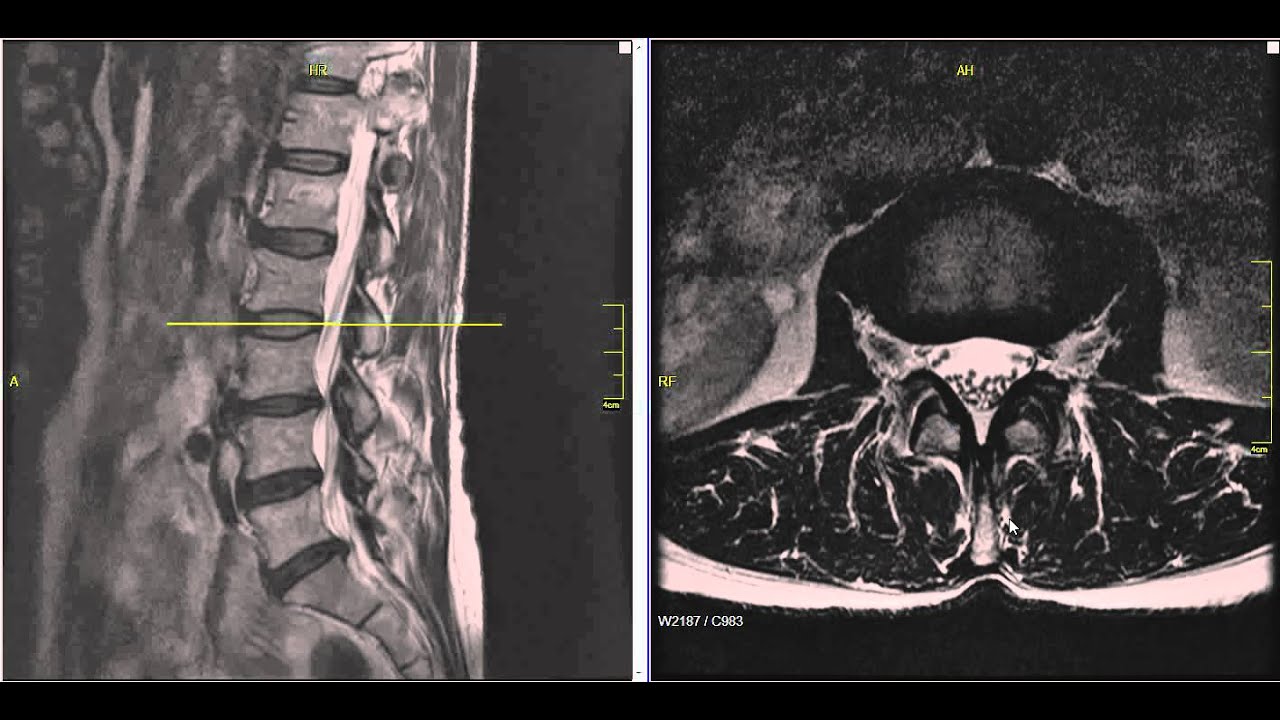
Free MRI Review.

. A herniated disc is frequently treated with nonsteroidal anti-inflammatory medication if the pain is only mild to moderate. If the diagnosis reveals mild to moderate injury IVVD treatment may include the administration of steroids and anti-inflammatory medications to reduce swelling and pain with confined rest. Magnetic resonance imaging MRI.
A slipped disc and ruptured disc are both other names for a herniated disc. Herniated disks are also called ruptured disks or slipped disks although the whole disk does not rupture or slip. CT scan generates cross section images of the spinal structures.
If the x-ray is being done to diagnose an injury care will be taken to prevent further injury. You will be asked to lie on the x-ray table in different positions. Getting X-rays helps rule out other causes of back or neck pain.
And an X-ray to locate the pressure on the spinal cord. The intervertebral disc space is typically defined on an X-ray photograph as the space between adjacent. Spinal disc herniation is an injury to the cushioning and connective tissue between vertebrae usually caused by excessive strain or trauma to the spineIt may result in back pain pain or sensation in different parts of the body and physical disabilityThe most conclusive diagnostic tool for disc herniation is MRI and treatment may range from painkillers to surgery.
A CT scanner takes a series of X-rays from different directions and then combines them to create cross-sectional images of the spinal column and the structures around it. 3 Telltale Signs of a L4-L5 Slipped Disc. Back pain is one of the most common causes for patients to seek medical care in both primary care and emergency setting.
Diagnosis The diagnosis of a C5 C6 bulging disc often includes a physical exam and imaging tests. Nerve root pain is usually classified according to the location of the affected nerve. A CT scan show the bones of your spine.
The best treatment method is through conservative physiotherapy physical therapy chiropractic and splinting night guards. X-ray MRI scan myelogram CT. More specialized tests include magnetic resonance imaging or myelogram which involves injecting a contrast dye into the spinal.
Otherwise known as Sciatica lumbar nerve pain is a combination of back and leg pain caused by nerve root inflammation in the lower backThe discomfort caused by lumbar nerve root inflammation is most severely felt in the section of the. Plain X-rays dont detect herniated disks but they can rule out other causes of back pain such as an infection tumor spinal alignment issues or a broken bone. An MRI uses radio waves a magnetic field and a computer to.
Another kind of herniation of the. A herniated disc is frequently treated with nonsteroidal anti-inflammatory medication if the pain is only mild to moderate. A herniated disk on the other hand results when a crack in the tough outer layer of cartilage allows some of the softer inner cartilage to protrude out of the disk.
Learn about degenerative disc disease sciatica and radiculopathy causes symptoms cervical lumbar thoracic buttock pain pain down leg diagnosis and treatment. And it includes tests such as an X-ray with dye contrast myelography MRI magnetic resonance imaging and CT scan computed tomography. It bulges usually in just one direction.
Have your primary doctor perform an MRI to be sure if the problem persists. The disc is not physically slipped. If you have serious persistent back pain caused by a bulging or.
It is the most common reason for workmans compensation and lost work hours and productivity. These tests will. Only the small area of the crack is affected.
The test is done in a hospital x-ray department or your health care providers office by an x-ray technician. Sometimes the herniation is so severe that a free fragment occurs meaning a piece has broken completely free from the disc and is in the spinal canal. The doctor may recommend physical therapy.
They may order imaging tests such as an X-ray or CT scan if they need more information about the condition of the spine and disks. Difficulty maintaining posture could also be caused by a slipped or bulging disc. The exam will generally include a neurological exam X-rays andor special imaging myelogram CT scan MRI to locate the source of spinal injury.
The doctor may recommend physical therapy. The most common and accurate imaging test for a suspected herniated disk is an MRI. Could you have a slipped or herniated disk.
More than 60 of people show evidence of disc degeneration at one or more levels on magnetic resonance imaging MRI. A myelogram to look for multiple slipped discs via X-ray after a dye is inserted into your spinal fluid. When no rupture or tear is present in a protruding disc it is considered a bulging disc.
Degenerative disc disease is the gradual deterioration of the discs between the vertebrae. An epidural steroid injection may be performed utilizing a spinal needle under X-ray guidance to direct the medication to the exact level of the disc herniation. X-ray helps in determining if there is any degeneration bony malformations fractures arthritis infection or tumors in the joints.
Actual X-ray cost depends on the provider the part of the body being X-rayed and the number of views taken. Imaging such as x-ray CT scan or MRI may be done as well as electromyography to measure nerve impulses in the muscles. A herniated ruptured or slipped disc means that a vertebral disc one of the soft pads of tissue that sit between each of the.
A true herniated disc also called a ruptured or slipped disc occurs when the disc annulus cracks or ruptures allowing the gel-filled center to squeeze out. Herniated disks can move into the space around your spinal cord and nerves and press on them. There are two broad categories namely.
In most cases the bones seen on X-ray are normal. An epidural steroid injection may be performed utilizing a spinal needle under X-ray guidance to direct the medication to the exact level of the disc herniation. MRI helps in assessing the involvement of any soft tissue and also helps in visualizing the discs nerve roots and spinal cord.
There is a broad range of potential etiologies for both. Pain that radiates down the leg is a symptom of sciatica. An estimated 200 billion dollars are spent annually on the management of back pain 1.
Symptoms of Slipped Disc in Dogs. A protruding disc is considered herniated when a rupture or tear is present. A pinched nerve in the neck often improves with simply a few days or weeks of rest.
Integrative alternative and complementary therapies - Health Professional Information NCI. September 6th 2017 Back Pain Amy Crowell. X-ray films are of some use but MRI assessment is the best means of confirming a slipped jaw disc diagnosis.

Lumbar Spine Image Medical Anatomy Radiology Imaging Medical Knowledge

Lumbar Spine Anatomy On Mri Magnetic Resonance Imaging Magnetic Resonance Imaging Magnetic Resonance Mri

Pin On Mri

Herniated Disc Spinal Disc Herniation Wikipedia Spinal Disc Herniation Disk Herniation Herniated Disc Exercise

Pin On Disc Disease

Pin On Bulging Disc

X Ray Stock Photos Royalty Free Images Vectors Video Intervertebral Disc X Ray X Ray Images

Pin On Back

Pin On Resonancia Ortopedia

Stem Cells For Degenerative Disc Disease Ddd Therapy Http Stemcellthailand Org Therapies Degener Degenerative Disc Disease Degenerative Disc Medical Anatomy
หมอ กายภาพบำบ ด ส ขภาพ

T2 Mri Lumbar Spine Sagittal View L4 5 Sever Disc Herniation With Spinal Stenosis Disc Degeneration At L4 L5 Also Lumbar Lordosis Spinal Stenosis Stenosis

Neck And Back Degenerative Spondylolisthesis Conditions Spondylolisthesis Lumbar Spinal Stenosis Spinal Degeneration

Mri Spine Anatomy Free Mri Lumbar Spine Sagittal Cross Sectional Anatomy Anatomy Images Mri Spines

Pin On Conditions

Prolapsed Cervical Disk Intervertebral Disc Pathology Nucleus Pulpusus Bulges Out Into A Weakend Area Of Annulus Fibr Herniated Disc Cervical Disc Herniated
Pin On Learning It S All Good

Herniated Lumbar Disc By Kris B Siemionow Md Leading Expert In Complex Spinal Surgery Disk Herniation Mri Spondylolisthesis

Mri Of Lower Lumbar Medical Radiography Radiology Imaging Medical Anatomy


